
Hair and Scalp tests
TESTING
1 min read
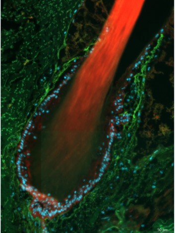
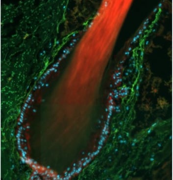
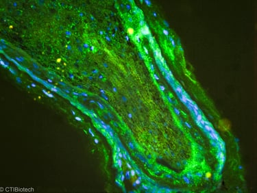
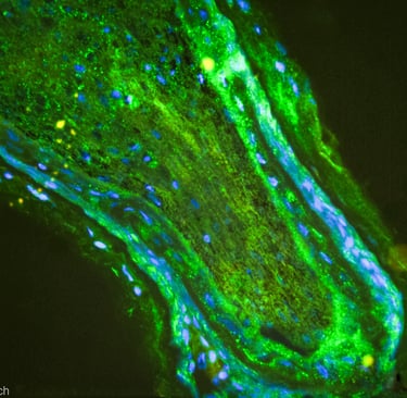
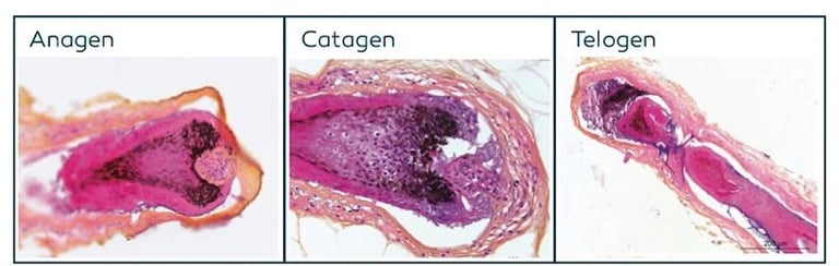
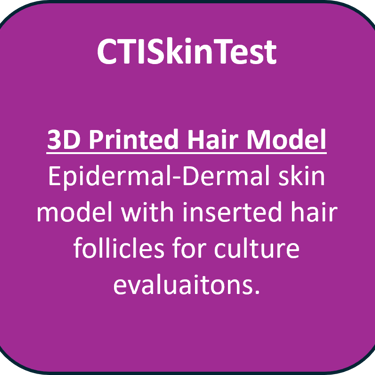
Few organisations carry out hair research and testing because it is extremely hard to do. At CTISKIN we have created protocols for individual hair follicles for short term culture, image analysis, and longer term studies. Scalp analysis is also possible.
Analysis can be made of:
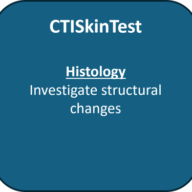
- the hair structure
-hair phasing
-hair growth
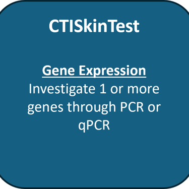
-follicular genes
-relation to the dermis and epidermis
-high resolution imaging


Click on the box to enlarge. For more information on the test contact us. We have extensive partners globally to help expand the range of tests we can offer. If you can't find a particular test, we probably have access to it.
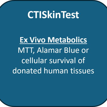
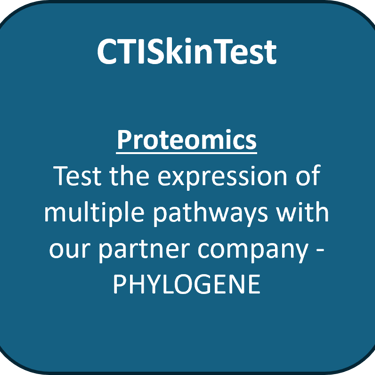
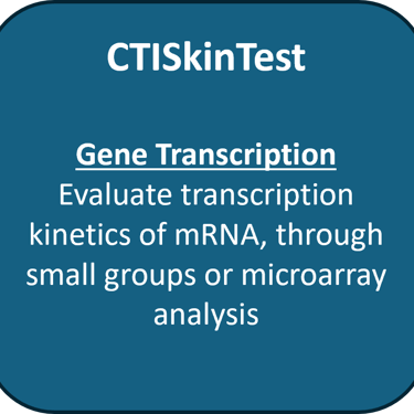
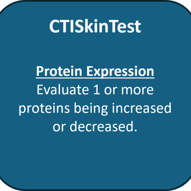
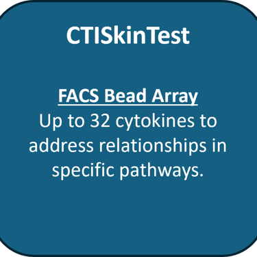
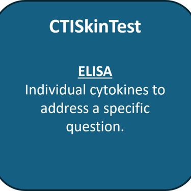
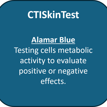
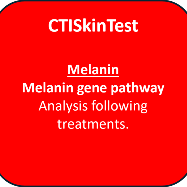
Tests we offer in this category:
Publications of relevance
Get In Touch
Want to discuss your project for testing?
Want to collaborate on research with us?
Want to buy cells and tissues for skin research and testing?
Contact us here!
Address
CTIBIOTECH
Bat A16, 5 avenue Lionel Terray
69330 Meyzieu
Lyon, France